The Brain Simulation Platform "Live Papers"
A biophysically detailed computational model of urinary bladder small DRG neuron soma

Authors: Darshan Mandge and Rohit Manchanda

Author Information: Computational Neurophysiology Lab, Department of Biosciences and Bioengineering, Indian Institute of Technology Bombay, Mumbai, India 400076

Corresponding author: Rohit Manchanda ( rmanch@iitb.ac.in )

Journal: PLOS Computational Biology,

Download URL: https://journals.plos.org/ploscompbiol/article?id=10.1371/journal.pcbi.1006293,

Citation: Mandge D, Manchanda R (2018) A biophysically detailed computational model of urinary bladder small DRG neuron soma. PLoS Comput Biol 14(7): e1006293. https://doi.org/10.1371/journal.pcbi.1006293,

DOI: https://doi.org/10.1371/journal.pcbi.1006293,

Licence: the Creative Commons Attribution (CC BY) license  applies for all files. Under this Open Access license anyone  may copy, distribute, or reuse the files as long as the authors and the original source are properly cited.

Abstract:
Bladder small DRG neurons, which are putative nociceptors pivotal to urinary bladder function, express more than a dozen different ionic membrane mechanisms: ion channels, pumps and exchangers. Small-conductance Ca2+-activated K+ (SKCa) channels which were earlier thought to be gated solely by intracellular Ca2+ concentration ([Ca\]i ) have recently been shown to exhibit inward rectification with respect to membrane potential. The effect of SKCa inward rectification on the excitability of these neurons is unknown. Furthermore, studies on the role of KCa channels in repetitive firing and their contributions to different types of afterhyperpolarization (AHP) in these neurons are lacking. In order to study these phenomena, we first constructed and validated a biophysically detailed single compartment model of bladder small DRG soma constrained by physiological data. The model includes twenty-two major known membrane mechanisms along with intracellular Ca2+ dynamics comprising Ca2+ diffusion, cytoplasmic buffering, and endoplasmic reticulum (ER) and mitochondrial mechanisms. Using modelling studies, we show that inward rectification of SKCa is an important parameter regulating neuronal repetitive firing and that its absence reduces action potential (AP) firing frequency. We also show that SKCa is more potent in reducing AP spiking than the large-conductance KCa channel (BKCa) in these neurons. Moreover, BKCa was found to contribute to the fast AHP (fAHP) and SKCa to the medium-duration (mAHP) and slow AHP (sAHP). We also report that the slow inactivating A-type K+ channel (slow KA) current in these neurons is composed of 2 components: an initial fast inactivating (time constant ~ 25-100 ms) and a slow inactivating (time constant ~ 200-800 ms) current. We discuss the implications of our findings, and how our detailed model can help further our understanding of the role of C-fibre afferents in the physiology of urinary bladder as well as in certain disorders.

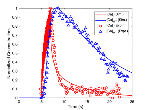
This Live Paper introduces interactively urinary bladder small-diameter DRG neuron soma, including modelling response to current clamps for action potentials, subthreshold potentials as well as the cytoplasmic and mitochondrial calcium transients.

Model Structure

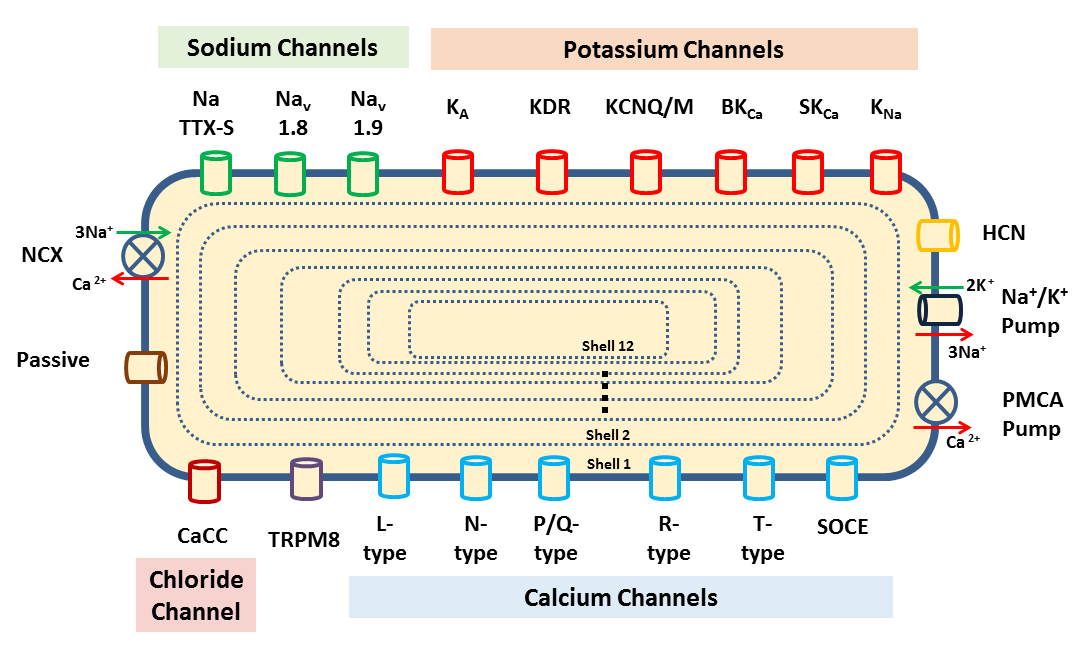
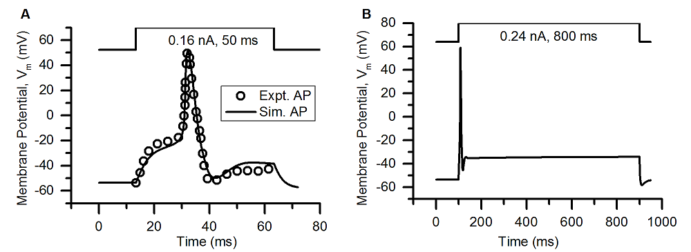
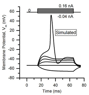
Fig. 1 Computational Model of Urinary Bladder Small DRG Neuron

Model response to Current Clamps

The captions in Fig. 2, Fig. 3 and Fig. 4 are linked to a Python Jupyter Notebook available in the Collab dedicated to this Live Paper through the Human Brain Project Collaboratory platform. The Jupyter Notebook allows to generate the simulation figures reported in the paper using the respective simulation conditions reported in figure captions and text.
More details on how to use the notebook and the results it allows to reproduce are reported inline with the code.


Fig. 2 AP Validation: [A] Single AP, [B] AP with a Long Duration Clamp


Fig. 3 Simulated Subthreshold Potentials and an AP in the model


Fig. 4 Comparing Simulated and Experimental Calcium Transients in the Cytoplasm (Red) and the Mitochondria (Blue).

Resources